Interproximal initial carious lesions diagnosed and monitored with Overjet AI
13
Anna Blake, RDH
USA
Product: Curodont Repair Fluoride Plus
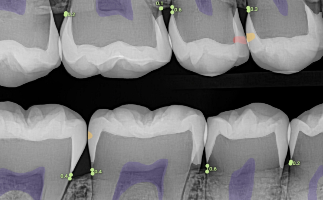
Before
Figure 1: Initial carious lesion on #4 mesial (D1), #5 distal (E2), and #30 distal (E1)
After
Figure 2: At the 6 month follow up, reduction in lesion size on #4 and complete reduction of lesion sizes on #5 and #30.
Patient Presentation and examination:
Personal information and relevant dental history: 17-year-old male, with a history of poor oral hygiene maintenance
Routine radiographic examination: Small radiolucencies were observed in the enamel of #4 (mesial), #5 (distal), and #30 (distal). This was confirmed using an artificial intelligence software (Overjet) (Figure 1)
Diagnosis:
#4 : Initial caries extending to the outer third of dentin (D1) on the mesial surface
#5 : Initial caries extending to the inner half of enamel (E2) on the distal surface
#30 : Initial caries extending to the outer half of enamel (E1) on the distal surface
Treatment:
The lesions were treated with Curodont Repair Fluoride Plus.
Oral hygiene instructions were reinforced.
Follow-up: At the 6-month follow-up post-treatment, the following observations were made, confirmed also with the AI software: (Figure 2)
The oral hygiene of the patient did not show much improvement
#5, #30: Reduction in the sizes of the treated lesions to E0, indicative of complete remineralization.
#4: Reduction in lesion size to E2, indicative of remineralization.
Take-away: Interproximal initial carious lesions, being asymptomatic, often escape detection. Routine radiographic examination, aided by AI interpretation, when possible, can help identify these lesions before they become cavitated. These can then be treated non-invasively with Curodont Repair Fluoride Plus, along with patient education on improving oral hygiene and diet, to help remineralize them.