Interproximal initial carious lesion adjacent to a restored tooth
11
Dr. William Moorhead
USA
Product: Curodont Repair Fluoride Plus
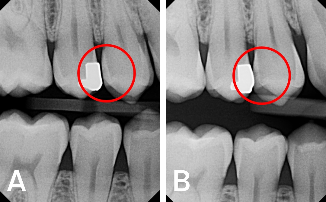
Before
Figure 1: Initial carious lesion on #5(distal), seen extending to the outer half of enamel (E1) on January 2022 (A) and found to have progressed to the inner half of enamel and DEJ (E2) on January 2023 (B)
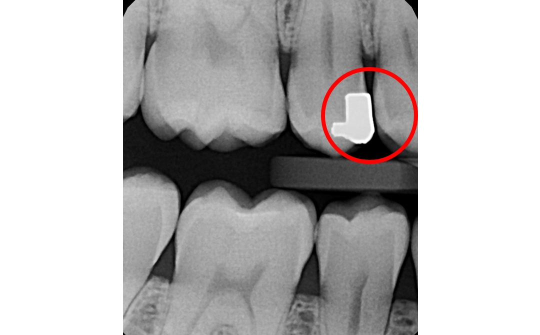
After
Figure 2: At the 5 month follow up, reduction in size and radiolucency seen.
Patient Presentation and examination:
Personal information: 26-year-old male
Routine radiographic examination:
A small radiolucency in the enamel of #5 (distal) was observed, which was adjacent to an old faulty amalgam restoration on the mesial surface of #4. (Figure 1A)
Initially kept on a ‘watch’, the radiolucency increased in depth in an x-ray taken a year later. (Figure 1B)
Diagnosis:
#5: Initial caries extending to the inner half of the enamel (E2) on the distal surface.
Treatment:
The lesion was treated with Curodont Repair Fluoride Plus in the same appointment.
Follow-up: At the 4-month follow-up, the following observations were made: (Figure 2)
Reduction in the size and radiolucency of the treated lesion to the very outer enamel, indicative of remineralization.
Take-away: Interproximal tooth surfaces adjacent to surfaces with faulty restorations, in this case an overhanging restoration, are at a risk of developing caries due to food entrapment and the difficulty in effective cleaning. In case of an inability to re-do the restoration correctly, such as when there is lack of patient consent, repeated follow ups to prevent and treat initial carious lesions on the adjacent proximal surface can be invaluable.